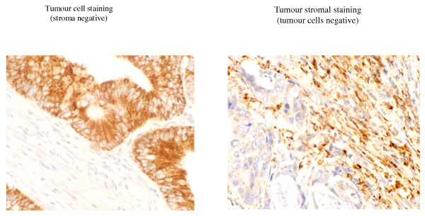
Figure 2.
Immunohistochemistry for APRIL in resected colorectal adenocarcinomas. Staining for APRIL was seen in the tumour cells (membrane and cytosol) and stroma (extracellular matrix and stromal cells) of colorectal adenocarcinomas. All combinations of tumour cell and stromal staining were seen. Tumour cell staining could be scored weak, moderate and strong. Examples show strong tumour cell staining and stromal staining.

