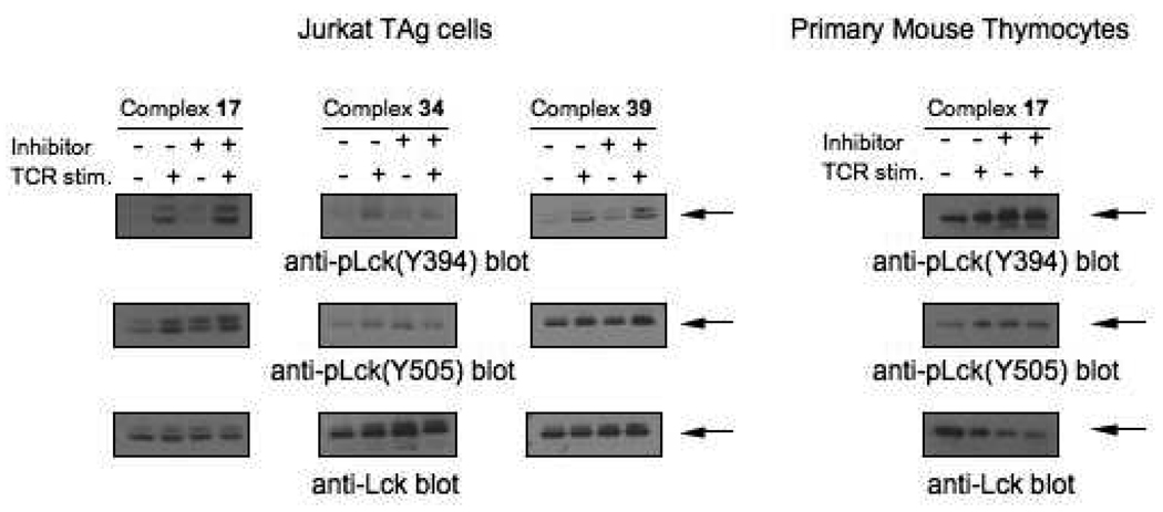
Figure 3.
Intracellular inhibition of LYP by gold complexes. (A) Top panels: anti-pLck(Tyr394) immunoblots of lysates of Jurkat TAg cells treated with 50 µM of complexes 17 and 34 and 25 µM of complex 39 (lanes 3 and 4 in each panel) or untreated (lanes 1 and 2 in each panel) and either left unstimulated (lanes 1 and 3 in each panel) or stimulated (lanes 2 and 4 in each panel) with C305 antibodies for 2 min. Middle panels: anti-pLck(Tyr505) blots of the same samples. Bottom panels: anti-Lck blot of the same samples. Arrows indicate the position of Lck (56 KDa) in each panel. (B) Top panel: anti-pLck(Tyr394) immunoblot of lysates of mouse thymocytes treated with 50 µM of compound 17 (lanes 3 and 4 in each panel) or untreated (lanes 1 and 2 in each panel) and either left unstimulated (lanes 1 and 3 in each panel) or stimulated (lanes 2 and 4 in each panel) with biotinylated anti-CD3 and anti-CD4, followed by cross-linking with streptavidin for 1.5 min. Middle panel: anti-pLck(Tyr505) blot of the same sample. Bottom panel: anti-Lck blot of the same sample.

