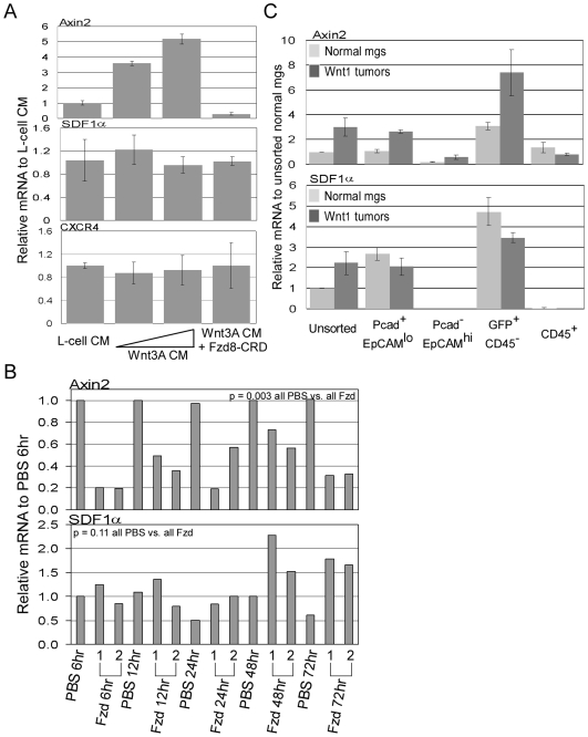
Figure 3. Determination of whether SDF1 is a Wnt signaling responsive gene.
A, qRT-PCR analysis of axin2, SDF1α, and CXCR4 transcript levels in normal mammary epithelial cells cultured in Wnt3A conditioned media. Triplicate plated cells were cultured in control L-cell media∶mammary epithelial cell culture media (1∶1 dilution), in Wnt3A conditioned media (1∶2 or 1∶1 dilution), and in Wnt3A conditioned media (1∶1) + Fzd8-CRD (10 µg/mL). Relative transcript levels were normalized to levels in L-cell media. Error bars represent standard deviations derived from triplicate plated cells. B, qRT-PCR analysis of axin2 and SDF1α transcript levels in Wnt1 tumors taken from tumor-bearing mice that were treated with Fzd8-CRD at various hours of post injection. Each time points contain samples from two independently FzD8-CRD treated mice numbered 1 and 2. Relative transcript levels were normalized to the level in the 6 hour PBS-treated tumor sample. C, qRT-PCR analysis of axin2 and SDF1α transcript levels in the sorted cell populations from Wnt1 tumors (dark bars) or from normal mammary glands (light bars). Cell populations were isolated from three independent Wnt1 tumors or from three independent pools of normal mammary glands (pools of glands from ∼20 female C57BL6 mice). Error bars represent standard deviations derived from the three independent tumors or pools of mammary gland sorts.

