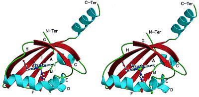
Figure 1.
Ribbons diagram of BjFixLH. The secondary structure elements are color-coded with α-helices as blue, β-sheets as red, and random coils as green. The atoms of the heme cofactor and the proximal histidine are shown as ball-and-stick models and are colored by their elements, with carbon as gray, nitrogen as blue, oxygen as red, and iron as green.

