
Figure 2.
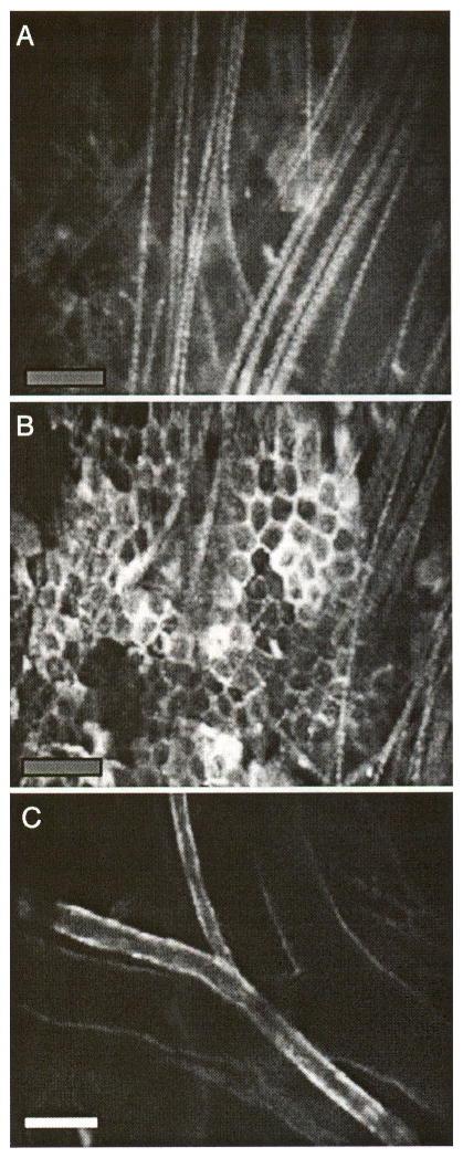
Optical sectioning by in vivo confocal microscopy. (A) Autofluorescent surface hair, (B) autofluorescent epidermal keratinocytes, and (C) an artery and vein pair in a plane deeper than the keratinocyte layer. The vessels are stained with Cy5.5-conjugated anti-PECAM-1 antibody that had been intravenously injected 16 hr before the images were taken. Bars indicate 100 μm.