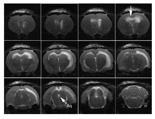
Figure 3.

T2-weighted MRI of animal with hydrocephalus induced by 10 KD dextran. Note the periventricular edema (arrow top row last figure from the left) in the corpus callosum and external capsule and the patent cerebral aqueduct (arrow labeled Aq). Note that the ventricular enlargement was asymmetric with the larger ventricle on the side of infusion.
