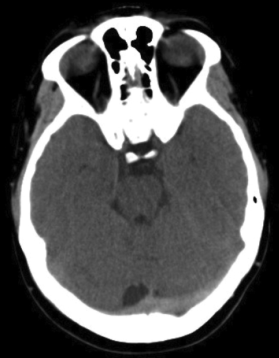
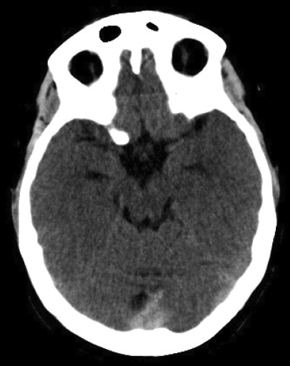
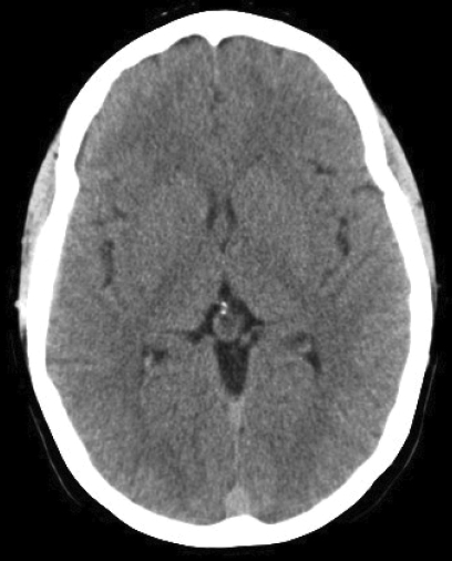
Figure 3.
Non-contrast axial CT scans (A–C) obtained at time of the onset of the patient’s symptoms were initially interpreted as normal. However, “rewindowing” makes cranial sinus thrombosis obvious. The only difference from her second CT scan (see figure 1▶) was the absence of right frontal venous hemorrhage. This example highlights the importance of meticulous examination of neuroimages to avoid missing diagnosis.


