Figure 2.
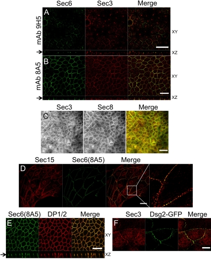
Exocyst localizes to desmosomes. (A) Polarized MDCK cells were colabeled with anti-Sec3CT and anti-Sec6(9H5) antibodies. Both en face (xy) and vertical (xz) confocal sections are shown, and the arrow denotes the position at which xy images were collected. The xz section demonstrates that endogenous Sec3 is enriched at sites on the lateral plasma membrane that are more basal than those labeled by anti-Sec6(9H5) antibodies. Bar, 20 μm. (B) Polarized MDCK cells were colabeled with anti-Sec3CT and anti-Sec6(8A5) antibodies. xy and xz confocal sections are shown. Note that Sec3 and Sec6 were colocalized at two distinct points of focal accumulation on lateral membranes. Bar, 20 μm. (C) Polarized MDCK cells were fixed in methanol and colabeled with anti-Sec3CT and a cocktail of anti-Sec8 mAbs (2E9, 5C3, 7E8, and 17A10). This method was required to visualize colocalization of Sec3 and Sec8 in polarized cells. Bar, 20 μm. (D) MDCK cells on collagen-coated coverslips were colabeled with anti-Sec15 and anti-Sec6(8A5) antibodies. Note that Sec15 and Sec6 were colocalized at puncta along contacting, but not noncontacting plasma membranes. The right-most panel shows a magnified view of the boxed area in the panel to its left. Bar, 20 μm. (E) Polarized MDCK cells were colabeled with anti-Sec6(8A5) and anti-desmoplakin 1/2 (DP1/2) antibodies. xy and xz confocal sections are shown. Note that DP1/2 and Sec6(8A5) were colocalized at two sites on the lateral membrane. Bar, 20 μm. (F) MDCK cells seeded on collagen-coated coverslips were transduced with Dsg2-GFP adenovirus and labeled with anti-Sec3CT antibody. Endogenous Sec3 colocalized with Dsg2-GFP-positive puncta at the plasma membrane. Bar, 10 μm.

