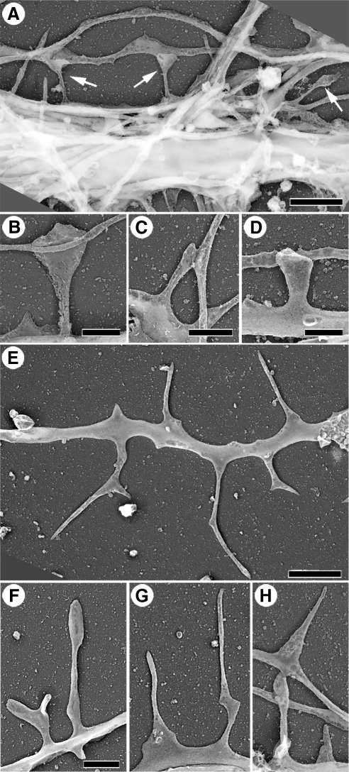
Figure 1.
Morphology of dendritic spines (A–D) and dendritic filopodia (E–H). Replica EM of nonextracted hippocampal neurons after 14 (A–D) or 10 (E–H) DIV. (A) Network of neurites with dendritic spines (arrows); two spines contact other neurites (axons). The cell body producing the major dendrite is located ∼20 μm away from the left margin of the panel. (B–D) Examples of mushroom (B), thin (C), and stubby (D) spines. (E) Distal region of a dendrite with polymorphic dendritic filopodia. (F–H) Examples of dendritic filopodia. Bars, 2 μm (A and E), 1 μm (B–D, F, and G), or 0.5 μm (H).

