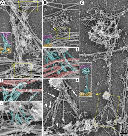
Figure 3.
Cytoskeletal organization of dendritic spines: base. EM of extracted neurons after 11 (A) or 14 (B and C) DIV shows undecorated (A and B) or S1-decorated (C) mushroom (A and C) or stubby (B) spines. The spine in A also may be considered stubby. Unlabeled insets show the smaller versions of the corresponding main panels with axons color-coded in purple, dendrites in yellow, and spines in cyan. Yellow boxes are enlarged in panels with corresponding numbers. Box 1, interaction of actin filaments (cyan) with axonal microtubules (red). Boxes 2 and 3, branched actin filaments (cyan) in the spine bases associate with dendritic microtubules (red). Box 4, linear actin filaments in the spine base have unbound ends (arrows) and are arranged in a delta-shaped configuration. S1-decorated actin filaments have a rough bumpy outline that distinguishes them from undecorated thin filaments (arrowheads). Bars, 0.2 μm (A–C).

