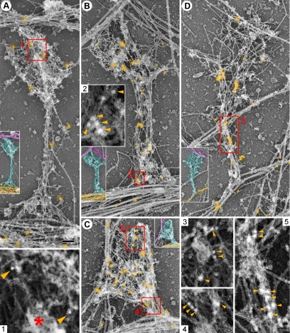
Figure 5.
Immunogold EM of cytoskeleton-associated proteins in spines. Unlabeled insets show the smaller versions of the corresponding main panels with axons color-coded in purple, dendrites in yellow, and spines in cyan. (A) N-cadherin. (B) Arp2/3 complex. (C) Capping protein. (D) Myosin II. Gold particles showing protein localization are highlighted in orange. Red boxes are enlarged in panels with corresponding numbers to show noncolorized gold particles (arrowheads). Box 5 shows a linear cluster of gold particles at the bottom. Bar, 0.2 μm.

