
Figure 7.
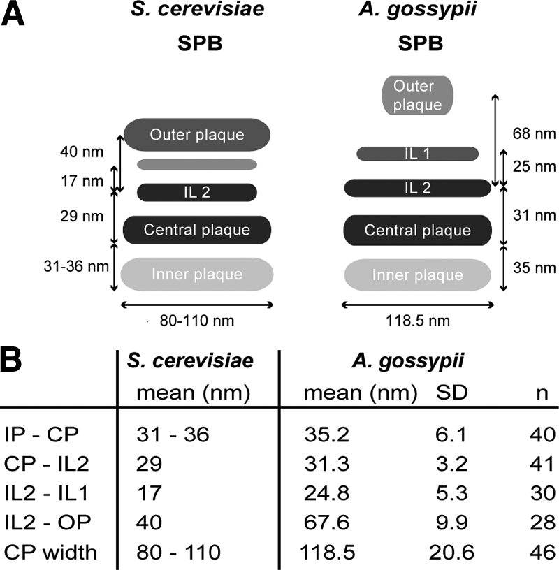
Comparison of A. gossypii and S. cerevisiae SPB structure based on EM analysis. (A) Schematic depicting the S. cerevisiae and A. gossypii SPB layers and distances between SPB layers. Although the CP and IL2 are similar in size and structure in A. gossypii and S. cerevisiae, the IL1 and OP of A. gossypii are considerably smaller and the spacing between those layers is increased compared with budding yeast. The A. gossypii OP appears amorphous rather than electron dense like in S. cerevisiae. (B) Quantitation of distances between A. gossypii SPB layers with SD and number of plaques used for the measurements. The data for S. cerevisiae SPB plaques were compiled from published work (Byers and Goetsch, 1974; Bullitt et al., 1997; O'Toole et al., 1999; Schaerer et al., 2001).