Figure 2.
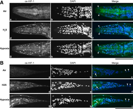
H2S exposure causes HIF-1 nuclear localization. Both H2S and hypoxia (0.5% O2) treatment induce HIF-1 nuclear localization throughout the animal. Images are stacks of all nuclei visible with DAPI staining. Animals were stained with anti-Ce-Hif-1 antibody (a, d, and g) and DAPI (b, e, and h) to visualize nuclei. Hypodermal nuclei (tailless arrows) and intestinal nuclei (tailed arrows) are clearly observed in the merged images (c, f, and i). (A) Anterior of the worm. (B) Posterior of the worm. Bars, 50 μm.

