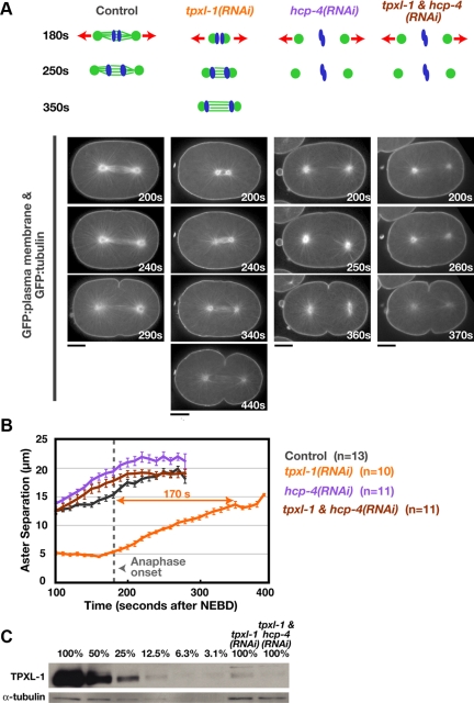
Figure 1.
TPXL-1 depletion introduces a delay between anaphase onset and the point when the asters achieve a normal extent of separation. (A) Schematics summarize the effects of each perturbation. Kinetochore microtubules, which resist cortical forces that pull on astral microtubules (red arrows), are absent in hcp-4(RNAi) embryos. Confocal images of embryos coexpressing GFP:β-tubulin and a GFP plasma membrane probe. Times are in seconds after NEBD. Bars, 10 μm. (B) Mean aster-to-aster distance, measured from the sequences in A, is plotted versus time in seconds after NEBD. Error bars are the SEM. Dotted line marks the mean time of anaphase onset in control and TPXL-1–depleted embryos. (C) Western blot of control, tpxl-1(RNAi), and tpxl-1 & hcp-4(RNAi) worms. Serial dilutions of the control lysate were used to quantify the amount of TPXL-1 in the RNAi samples (percentage of amount in 100% control indicated above each lane). RNAi of tpxl-1 alone and tpxl-1 & hcp-4 reduced TPXL-1 levels to 7.4 and 5.6% of that in controls, respectively.

