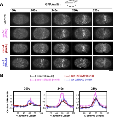
Figure 6.
GFP:Anillin accumulates on the equatorial cortex with normal timing after depletion of SPD-1, ZEN-4, or AuroraBAIR-2. (A) Spinning disk confocal optics were used to image the cortex in control (n = 46), spd-1(RNAi) (n = 10), zen-4(RNAi) (n = 13), and air-2(RNAi) (n = 10) embryos expressing GFP:Anillin. Images are maximum intensity projections of four cortical sections collected at 1-μm z intervals. (B) The mean postanaphase accumulation of cortical GFP:Anillin was quantified as a function of embryo length (as described for Figure 3) for control, spd-1(RNAi), zen-4(RNAi), and air-2(RNAi) embryos at the indicated time points after NEBD. Error bars are SEM. Bar, 10 μm.

