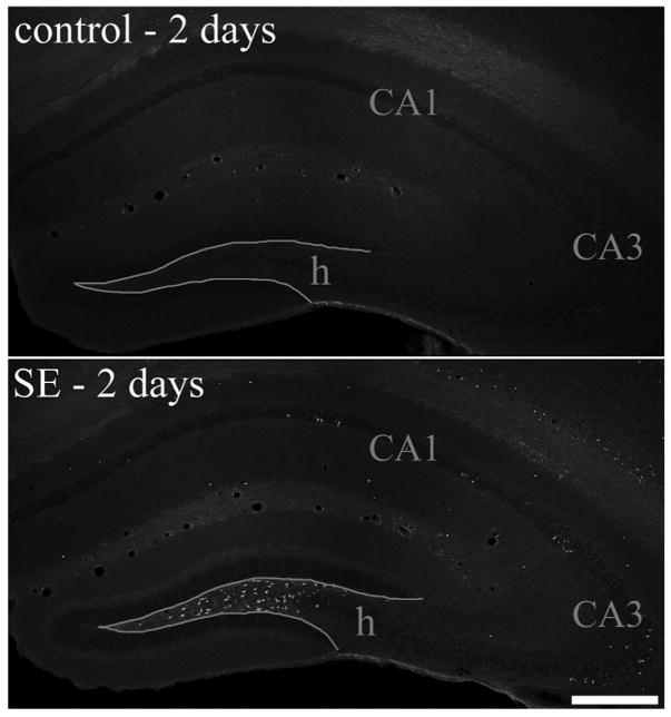
FIGURE 4.
Fluoro-Jade B staining of hippocampal sections from a control animal and pilocarpine-treated animal sacrificed 2 days after status epilepticus (SE). Fluoro-Jade B staining was absent from control animals (top), whereas in pilocarpine-treated animals large numbers of labeled cells were present in the dentate hilus (h) and scattered cells were labeled throughout the CA1 and CA3 pyramidal cell layers. Scale bar = 300 μm. [Color figure can be viewed in the online issue, which is available at www.interscience.wiley.com.]

