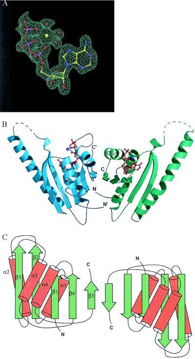
Figure 1.
(A) A sample of the electron density map for ATP in the ATP-binding pocket of MJ0577. The multiwavelength anomalous diffraction-phased electron density map at 1.8-Å resolution is contoured at 1 sigma, with the current model displayed for comparison. A Mn ion and three waters bound to ATP are also shown. (B) Ribbon diagram of the crystal structure of the MJ0577 dimer drawn by using molscript (29) and raster3d (30). The secondary structure assignment is based on dssp (31). Dashed lines represent loops not modeled in the structure. The N and C termini of each monomer are labeled. (C) Schematic structure, where β-strands and α-helices are represented by arrows and cylinders, respectively.

