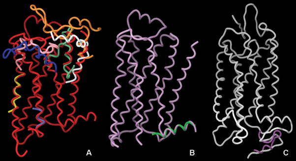
Figure 2.
Structural alignment of the NMR coordinates of GPCR portions deposited in the PDB and the crystal structures of the and bovine rhodopsin. Panel A: superimposition of the β2-AR crystal structure (2RH1, red) with the NMR-derived structures of the N-termini of PTH1 (1BL1–white) and CCK1 (1D6G, orange), EL1 of S1P4, (2DCO, aquamarine), IL3 of CB1 (1LVQ, light blue), TM6 of Ste2pR (1PJD, yellow), and EL3 of CCK1 (1HZN, pink) and CCK2 (1L4T, dark blue). Panel B: superimposition of the crystal structure of the β1-AR (2VT4, plum) with the NMR-derived structure of H8 of the same receptor (1DEP, green). Panel C: superimposition of the crystal structure of rhodopsin (2HPY, white) and the NMR-derived structure of the C-terminus of the same receptor (1NZS, blue purple). Pictures prepared with Maestro 8.0.308, Schrodinger.

