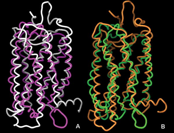
Figure 3.
Superimposition of the crystal structures and the NMR-based three-dimensional models of bovine rhodopsin. Panel A: Superimposition of the X-ray structure (1GZM, white) and the NMR-based model of the ground state (1LN6, purple) receptor. Panel B: Superimposition of the X-ray structure of opsin in its G-protein-interacting conformation (3DQB, orange) and NMR-based model of Meta II rhodopsin (1JFP, green). Picture prepared with Maestro 8.0.308, Schrodinger.

