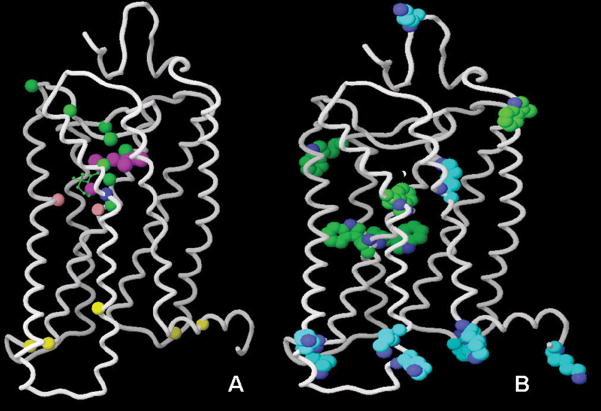
Figure 5.
Panel A: Positions of the ligand atoms and residues labeled in order to study the activation-related conformational changes of rhodopsin. The retinal atoms (C5, C9, C11, C13, C14, C15, C19, C20) are in purple; residues 114, 118, 121, 178, 188, 191, 196, and 268 are in green; residue 265 is in blue; residues 122 and 211 are in pink; the residues mutated to Cys to be labeled with trifluoroethylthio groups are in yellow. For simplicity, only one carbon of the residue backbone is shown. Panel B: All Trp and Lys residues of rhodopsin. Picture prepared with Maestro 8.0.308, Schrodinger.

