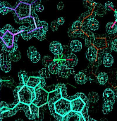
Figure 2.
A view of a hexahydrated magnesium ion (in green) bridging three DNA duplexes with the corresponding electron density (2Fo-Fc) map at 1.6 Å resolution contoured at 1.5σ. Hydrogen bonds between the Mg-coordinated water molecules (shown in red) and base or backbone donor/acceptor atoms from three helices (shown in blue, orange, and magenta) are drawn as yellow lines. The figure was prepared with o 5.10 (10).

