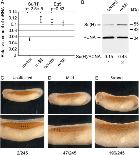
FIGURE 2.
α-SE morpholino derepresses endogenous Su(H) mRNA and causes segmentation defects. (A,B) Xenopus embryos at the two-cell stage were injected with 2 pmol of control or anti-Su(H) EDEN morpholino (α-SE) in each blastomere. They were allowed to develop until the tailbud stage (24–26). (A) RNAs were extracted and the amounts of Su(H) and Eg5 mRNA, relative to the amount of ODC mRNA, were measured by RT-qPCR. Shown are the medians, ±s.e., of 15 embryos for each condition. Student tests were used to compare control and α-SE morpholinos, and P-values are indicated above the bars. (B) Proteins were extracted from pools of five embryos, and one embryo equivalent was submitted to a Western analysis using antibodies directed against Su(H) and PCNA as a loading control. Positions of molecular weight markers are indicated on the right. (C–E) Two-cell embryos were injected in one blastomere with 2 pmol of anti-Su(H) EDEN morpholino (α-SE) and allowed to develop until tailbud stage. They were fixed and stained with myotome-specific 12/101 monoclonal antibody. C, D, and E are representative photographs of an unaffected embryo, an embryo with a progressive loss of segmentation toward the posterior extremity, and a nonsegmented embryo, respectively. Lower panels are higher magnifications of upper panels. In C and D, lower panels, stars show successive somites. The number of embryos that fell within each class in one representative experiment is given on bottom.

