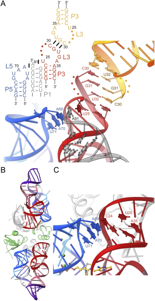
FIGURE 4.
Structural plasticity of P3. (A) Schematic secondary structure and cartoon representation of the P3–L5 region of the riboswitch in its crystalline context, in the structure given by Edwards and Ferré-D'Amaré (2006). Interactions formed with symmetry-related molecules in the crystal (gray and yellow) are shown. Note intercalation of A91 from a symmetry-related molecule between U25 and A69. (B) Cartoon representation of the new E. coli thiM aptamer domain crystal structure. The two RNA molecules in the asymmetric unit are colored as in Figure 1. (Gray) Cognate U1A-RBDs, (green) two additional U1A-RBDs that do not make specific RNA-binding interactions. (C) Detail of P3–L5 interaction in the new crystal structure. Note extrusion of C24 from P3, and its stacking on A69.

