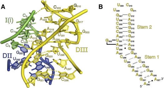
FIGURE 8.
Domain III. (A) Cartoon rendering of the internal loop and S-turn of DIII. Hydrogen bonds to domains 1 and 2 and those within DIII are shown. The strands are color-coded by secondary structural subdomain, as shown in Figure 1A. (B) Secondary structure diagram of DIII. Note that G300 is shown as a bulged nucleotide.

