Figure 2.
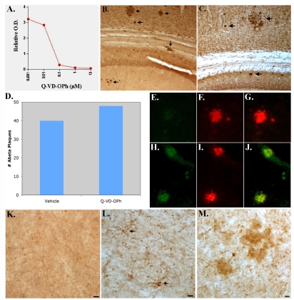
The effects of chronic administration of Q-VD-OPh on Aβ deposition, caspase-7 activation, and the caspase-cleavage of tau in TgCRND8 mice. A) Cell-free inhibition of caspase-7 activity by Q-VD-OPh. Data display a concentration-dependent inhibition of caspase-7 cleavage of the substrate AMC-DEVD-pNA in a cell-free system consisting of active human recombinant caspase-7. The calculated IC50 of Q-VD-OPh was determined to be approximately 48 nM. B-D) Three month-old TgCRND8 mice were injected 3 times a week for 3 months with 10 mg/kg of Q-VD-OPh (B) or vehicle (C). Data are representative of 3 independent experiments utilizing the monoclonal antibody to Aβ (clone 6E10, 1:400) and indicate the presence of extracellular Aβ deposits in the cortex and hippocampus of both animals (arrows). Panel D depicts semi-quantitative analysis of the number of Aβ plaques for vehicle or following Q-VD-OPh-treatment. E-J) Double-immunofluorescence images with active caspase-7 in green and 6E10 in red. (E-G) represent animal treated with Q-VD-OPh indicating that while caspase-7 activation was prevented (E), there was no apparent effect on plaque formation (F). Panel G represents the overlap image for Panels E and F. (H-J) Identical to above except panels represent a control animal treated with vehicle. In this case both active caspase-7 (H) and plaque labeling (I) are evident. (K-M) Prevention of the caspase-cleavage of tau following treatment with Q-VD-OPh. (K) Represents an animal treated with Q-VD-OPh, whereas Panels L and M represent vehicle controls following staining with the TauC3 antibody. No apparent staining was observed in the treated animal, while the presence of caspase-cleaved tau was evident in astrocytes (arrows, L) and extracellular plaques (M) in the vehicle controls. Scale bars represent 10 μm.

