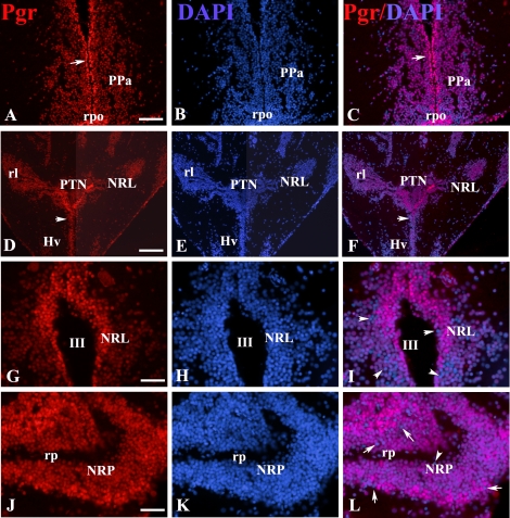
FIG. 10.
Representative immunoreactivity in the forebrain of an adult female zebrafish at the level of the anterior preoptic area (A–C), hypothalamus (D–I), and caudal line (J–L) for Pgr (red; A, D, G, and J) and DAPI (blue; B, E, H, and K). Superimposed images (C, F, I, and L) are shown. Arrows in A, C, D, and F indicate that the majority of the nuclei located in the periventricular layers have stronger Pgr immunostaining than those in the parenchyma. Pgr-negative nuclei were indicated by arrows in I and L. NPT, nucleus posterioris tuberis; NRL, nucleus recessus lateralis; NRP, nucleus recessus posterioris; PPa, anterior preoptic region; rpo, preoptic recess; III, third ventricle; rl, lateral recess; rp, posterior recess. Bars = 60 μm (A–C), 120 μm (D–F), or 30 μm (G–L).

