FIG. 2.
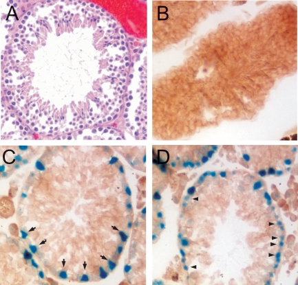
VRK1 is expressed in the basal layer of the seminiferous tubule in both Sertoli cells and spermatogonia. A) Representative tubule within a GT12/GT12 testis, following formalin fixation and H&E staining, is shown. The GT12/GT12 tubules exhibit wild-type morphology and cellular content. B–D) Frozen sections of testes dissected from 11-wk-old mice were assayed in situ for β-gal activity. A wild-type tubule (B) and GT12/GT12 tubules (C and D). Arrows shown in C point to some of the β-gal+ Sertoli cells. Arrowheads shown in D highlight some of the β-gal+ spermatogonia. Original magnification ×10.

