FIG. 2.
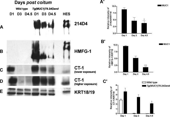
Human MUC1 expression in early pregnancy series in human MUC1 transgenics. Endometrial extracts from Days 1, 3, and 4.5 postcoitum (D1, D3, and D4.5, respectively) from both wild-type and human MUC1 transgenics were analyzed by Western blotting as described in Materials and Methods. Human MUC1 detection was with either 214D4 (A) or HMFG-1 (B) monoclonal antibodies specific for human MUC1. No signal was observed for either antibody in extracts from wild-type mice. Expression of human MUC1 is reduced, but it persists during early pregnancy. C) Detection of human and mouse MUC1 by CT-1 antibody. Almost complete loss of MUC1 expression is detected in the wild type, whereas MUC1 expression persists at Day 4.5 in the transgenics, reflecting the presence of the human MUC1. D) A longer exposure of samples shown in C. E) Blots were reprobed with antibody specific for mouse cytokeratin 18/19 (KRT 18/19), epithelial cell markers serving both as a load control and to control for changes in total epithelial cell populations during pregnancy. Total protein extract from HES cells was used a positive control. Bar graphs in A′, B′, and C′ represent densitometric analyses to quantify MUC1 expression normalized to KRT18 of the above data provided as mean ± SD values in each case. *P < 0.05 relative to human MUC1 Day 1; ***P < 0.001 relative to human MUC1 Day 1.

