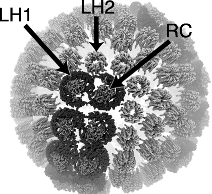
Figure 1.
Patch of the chromatophore from purple photosynthetic bacteria Rhodobacter sphaeroides showing the placement of LH2 and LH1-RC complexes as determined from atomic force microscopy and computational modeling (Ref. 5). Pools of LH2 antenna complexes are seen to surround the LH1-RC core complexes; lipids are not shown. As the primary antennae of the chromatophore most of the intercomplex excitation transfer occurs between LH2s.

