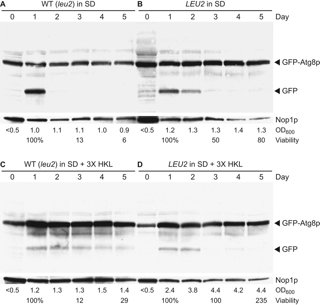
Figure 12.
Induction of autophagy during chronological aging. Control (WT) and LEU2 (YAA1) strains were transformed with plasmid pCuGFPAUT7(416) (Kim et al., 2001), which expresses a GFP-Atg8p fusion protein that is proteolytically processed in an autophagy-dependent manner to yield GFP. Transformants were grown in synthetic dextrose minimal medium containing standard amounts of the essential amino acids histidine, lysine, and leucine (SD) or containing three-fold increased amounts of the same supplements (SD + 3X HKL) as described in Table 2. Protein lysates from cells collected on days 0–5 were analyzed by western blotting using a GFP-specific polyclonal antibody (see Materials and Methods). Blots were reprobed with monoclonal antibody directed against the nucleolar protein Nop1p, which served as a loading control. Cell density (OD600) and percent viability values are shown below the lane corresponding to the day on which data were collected.

