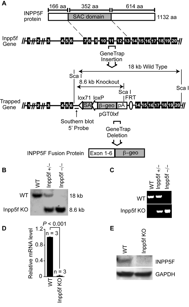
Figure 1. Inactivation of Inpp5f.
(A) Schematic representation of Inpp5f protein (top) and gene structure. The wild type and gene trap alleles are shown; exons are represented by black boxes. The sizes of the expected restriction fragments recognized by the Southern probe (indicated) are shown. (B) Southern blot of adult mice tail DNA resulting from a cross between Inpp5f+/− heterozygotes. (C) PCR genotyping of offspring resulting from a cross between Inpp5f+/− heterozygotes. (D) Real-time quantitative PCR of mRNA from adult wild-type and Inpp5f−/− hearts shows the absence of Inpp5f mRNA in the knockout hearts. (E) Western blot of wild-type and Inpp5f−/− adult heart tissue shows loss of Inpp5f protein in mutant hearts.

