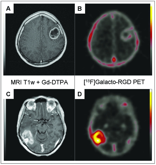
Fig. 1.
Examples of patients with glioblastoma multiforme of the left frontal lobe (A, B) and right parieto-occipital lobe (C, D). Note the intense peripheral enhancement in the gadolinium-DTPA–enhanced MRI scans in both tumors (A, C). However, the tumor in B shows only very faint tracer uptake in the [18F]Galacto-RGD PET (maximum standardized uptake value [SUVmax], 1.2), whereas the tumor in D demonstrates substantially more intense [18F]Galacto-RGD uptake (SUVmax, 2.8).

