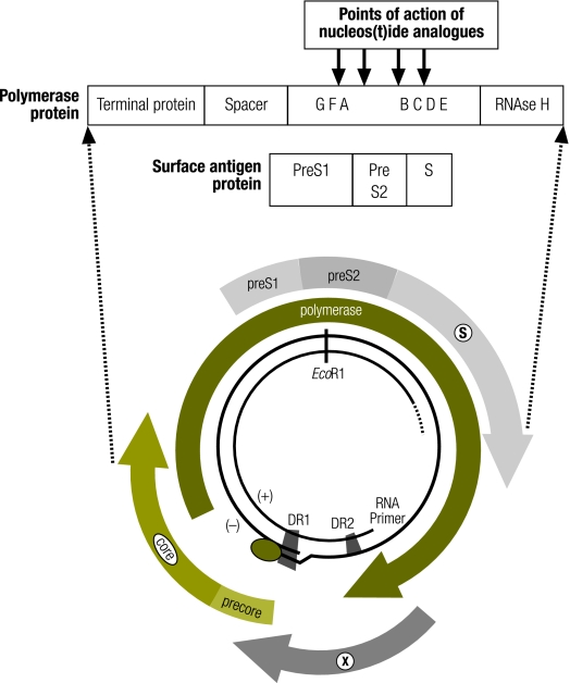
Fig. 1.
Genome structure of the hepatitis B virus showing overlapping reading frames of the polymerase and surface antigen
DR1, direct repeat sequence 1; DR2, direct repeat sequence 2; EcoR1, the cut site of the restriction endonuclease EcoR1 derived from E. coli; X, X gene encoding the HBV X protein; PreS1 and PreS2, large envelope proteins; S, the small envelope protein.

