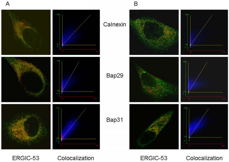
Figure 2. Colcalization images of ERGIC-53, a marker of the ER/Golgi intermediate compartment with calnexin (an ER marker), Bap29 and Bap31.

A) 37°C. All proteins were labeled with primary antibodies of different species or isotypes, followed by specific fluorescent anti-Ig. See ‘Methods’ for details. The left panels show the overlays of images of ERGIC-53 in red and the other proteins in green. The right panels are scatter plots of the pixels above a threshold that are both red and green. For ideal colocalization pixels are distributed along a diagonal. The spread away from but parallel to the diagonal is characterized by Pearson's coefficient. Pixels off the diagonal are due to bleed-through of one color into the other channel. It can be seen that there is excellent colocalization of ERGIC-53 and Bap31 (lower panels), and good colocalization of ERGIC-53 with calnexin and Bap29. B) Colocalization after cells were incubated at 15 °C to block recycling of ERGIC to the ER. There is still a significant fraction of Bap31 colocalized with ERGIC-53, indicated by the number of points distributed around the diagonal. There is little colocalization of calnexin or Bap29.
