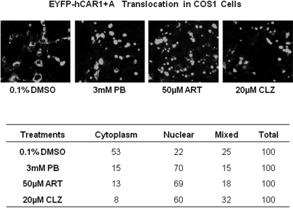
Fig. 5.
Translocation of EYFP-(hCAR1+A) in COS1 cells after treatment with known hCAR activators. COS1 cells were transfected with EYFP-(hCAR1+A) as described under Materials and Methods, and treated with 0.1% DMSO, PB (3 mM), ART (50 μM), or CLZ (20 μM). After 24 h of treatment, cells were subjected to confocal microscopy analysis. A, representative localization of EYFP-(hCAR1+A) in each treatment group. B, for each treatment, 100 EYFP-(hCAR1+A) expressing cells were calculated and categorized as cytoplasmic, nuclear, or mixed (cytoplasmic + nuclear) localizations.

