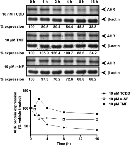
Fig. 8.
TMF fails to stimulate AHR protein degradation. Huh7 cells were treated with vehicle (DMSO), 10 nM TCDD, 10 μM TMF, or 10 μM α-NF for the indicated times. Total protein was harvested and used to assess AHR protein expression. Fifty micrograms of lysate were resolved by SDS-PAGE, transferred to PVDF membrane, and probed for AHR and the loading control β-actin by use of appropriate antibodies. Immune-reactive bands were visualized through autoradiography and quantified by γ-counting. Data represent percentage of AHR expression relative to vehicle-treated control.

