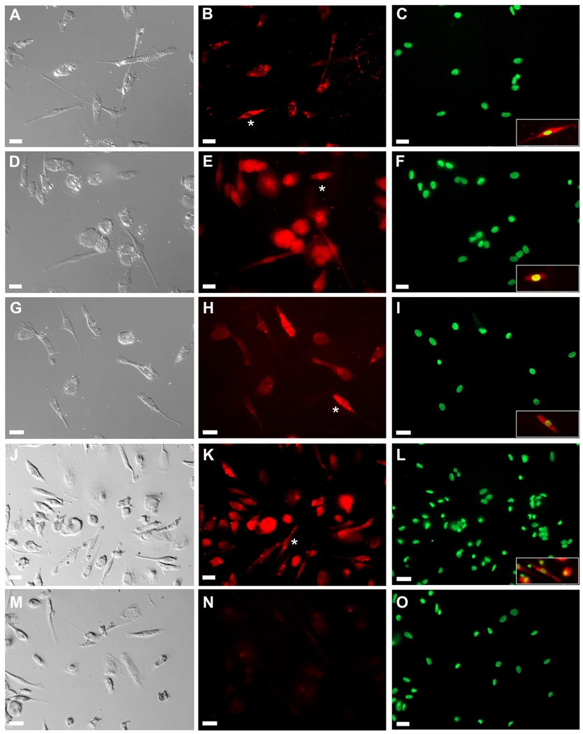
Figure 1. Morphological and immunohistochemical analysis of fibroblasts cultured from PB.
Morphological identification of fibroblasts was based on DIC images (Panels A, D, G, J and M). Cells were then stained with antibodies to SMA (Panel B), collagen type I (COL-1) (Panel E), vimentin (Panel H) or 5B5 (prolyl 4-hydroxylase; Panel K). Analysis showed that all cultured cells with morphological characteristics of fibroblasts expressed markers associated with this cell type. Panel N shows control staining with secondary antibodies only. Panels C, F, I, L and O show nuclear staining with Hoechst dye. Insets in Panels C, F, I, L and O show superimposition of antibody staining (red) and nuclear staining (green) for cells indicated by asterisks in Panels B, E, H, and K. Bar = 25 m.

