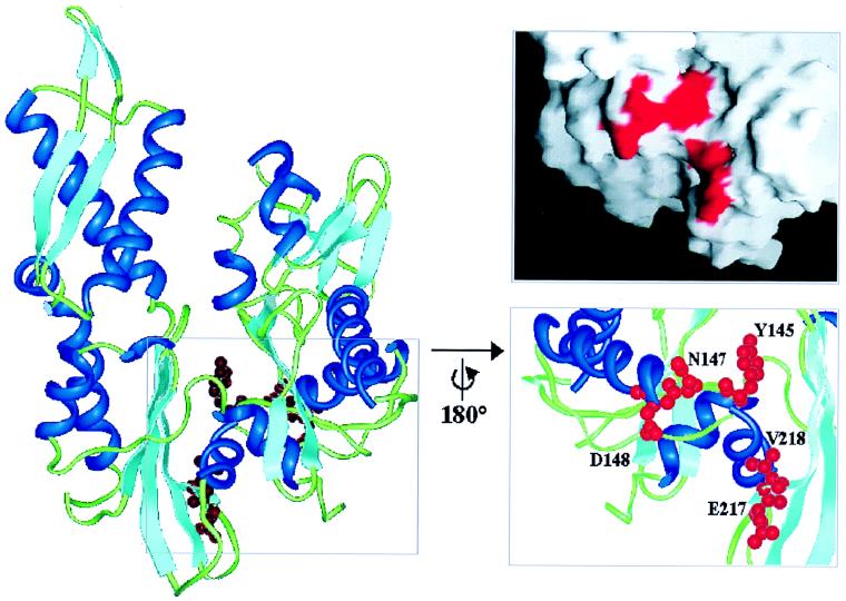
Figure 2.
Standard view of the structure of the DnaK ATPase domain within the DnaK–GrpE complex (PDB entry 1DKG) (21), showing the location of the mutated channel residues Y145, N147, D148, E217, and V218. α-helices are shown in dark blue, β-sheets in light blue, loops in green, and the side chains of the mutated residues in red. The marked region was rotated by 180° about the vertical axis. Residues Y145, N147, D148, E217, and V218 are shown as ball and stick models. The figures were prepared with insight II (Micron Separations, San Diego). The surface was created by using the program grasp (29), with mutated residues marked in red.

