Fig. 1.
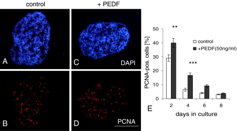
PEDF (50 ng/ml) increases proliferation in retinal spheroids, as identified by PCNA staining. Cryosections of 2-day-old spheroids were double-stained with DAPI (a, c) and with PCNA (b, d). e shows quantification of data. Note that sizes of sections do not reflect true volume size of spheroid (see “Materials and methods”). Each data point represents the mean ± SD of multiple sections (n = 6). **P < 0.001; ***P < 0.0001. Scale bar, 100 μm

