Fig. 3.
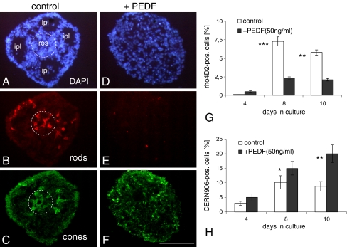
PEDF (50 ng/ml) decreases general spheroid tissue differentiation (shown by DAPI in a, d) and differentiation of rod photoreceptors (b, e) and increases number of cone photoreceptors (c, f). The IPL-like areas (ipl in a) and rosettes (ros, broken circles in a–c) are absent in PEDF-treated spheroids. Cryosections of 8-day-old spheroids were triple-stained with the rod-specific antibody rho4D2 (red; b, e) and the red and green cone-specific antibody CERN-906 (green; c, f). The percentage of rho4D2- and CERN906-positive cells was calculated in relation to DAPI-positive cells (g, h). Each data point represents the mean ± SD of multiple sections (n = 6). **P < 0.001; ***P < 0.0001. Scale bar, 100 μm

