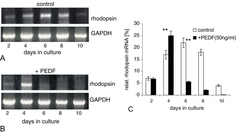
Fig. 4.
Temporal expression of rhodopsin mRNA in rosetted spheroids (a) or in the presence of 50 ng/ml PEDF (b), as analyzed by semi-quantitative RT-PCR at different culture days; the percentage of rhodopsin mRNA was calculated in relation to expression of GAPDH mRNA (c). Note the strong decrease of rhodopsin mRNA in the presence of PEDF (further see text). **P < 0.001

