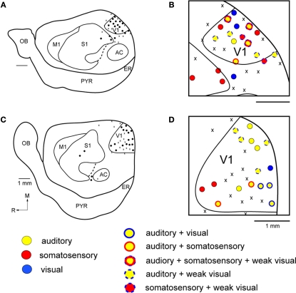
Figure 3.
Functional Organization of V1 in Gnat−/− Mice. Reconstructions of multi-unit electrophysiological recordings from 2 Gnat−/− mice are shown in which boundaries of primary sensory areas, as determined by myeloarchitecture, are denoted by black lines and recording sites are denoted by black dots indicating the location of recording sites (A,C). Enlargements of the recording sites in visual cortex are shown in which recording sites are denoted by color coded dots indicating the sensory modality mapped to that location (B,D). Unlike normal animals, in which neurons in V1 respond solely to visual stimulation, in Gnat−/− mice neurons at only a few recording sites responded only to visual stimulation. At most other sites in which neurons were responsive, they responded to auditory stimulation, somatosensory stimulation or some combination of modalities of stimulation. Medial is to the top and rostral is to the left in all images. X = No Response. Scale bars = 1 mm.

