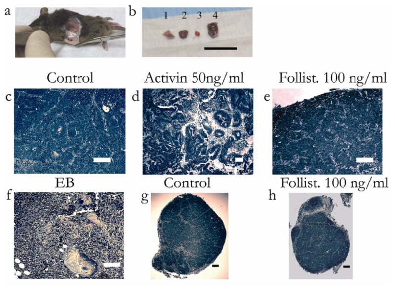
Figure 5.

In vivo differentiation of control, activin, and follistatin-treated cells. Day 10 gel cultured embryonic stem cells were mixed with NIH3T3 fibroblasts and subcutaneously implanted in syngeneic mice, for another 14 days. Conditions were, control (gel culture), activin (50ng/ml), and follistatin (100 ng/ml), and embryoid body control. (a) Encapsulated superficial mass demonstrated after incision. (b) Day 24 tissue masses after resection were embryoid body (1), gel culture (2), follistatin (100 ng/ml) (3), and activin (50 ng/ml) (4). Note that activin-treated cells resulted in the largest mass. Bar = 1 cm. H&E (Hematoxlin and Eosin) staining of implanted tissue masses. (c) Staining of gel culture demonstrates uniform epithelial-like cells with spindle-like septae. Bar = 100 μm. (d) Activin-treated cells, demonstrate heterogeneous, thick tubule-like structures, and mesenchymal elements. Bar = 100 μm. (e) Staining of follistatin-treated cells demonstrates uniform cords of epithelial cells separated by septae. Bar = 100 μm. (f) EB cells demonstrate heterogeneous tissue with skin, cartilage, and mesenchymal elements. Bar = 100 μm. (g) Low magnification image of the encapsulated gel culture mass. Bar = 200 μm. (h) Low magnification image of encapsulated follistatin-treated mass. Bar = 200 μm.
