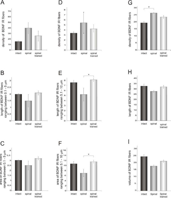
Figure 10.
BDNF immunoreactivity in processes and fibers of motor nuclei in the L3/L4 segments of the spinal cord of the intact, spinal and spinal trained rats labeled with the Santa Cruz antibody. The left and central columns present data analyzed with a skeletonization technique (Image-Pro Plus software). Density (A), length (B) and area (C) occupied by BDNF immunoreactive (IR) processes and fibers ranging in length from 0.1 to 5 μm (mean ± SEM). Central column: density (D), length (E) and area (F) occupied by BDNF IR processes and fibers ranging in length from 5.1 to 10 μm (mean ± SEM). Six intact, five spinal and six spinal trained rats were used and at least three sections per animal were analyzed. The density of BDNF IR processes tended to be higher in the spinal than in intact animals and locomotor training tended to normalize it (A, D). The mean length of BDNF IR processes and the area occupied by them (B, E, C, F) were lower in the spinal than in the intact animals and training also normalized them (p < 0.02, one-way Kruskal-Wallis test). Asterisk corresponds to p < 0.05 (Dunn post-hoc test). Right column: density (G), length (H) and volume (I) of BDNF IR processes and fibers traced with the aid of Neurolucida software in the area of 50 000 μm2 in the ventro-lateral nucleus. One section per animal was analyzed. Higher density of the BDNF IR network of processes and fibers observed in the spinal animals (G) was composed of shorter and thinner elements than in the intact and spinal trained animals (p < 0.05, one-way Kruskal-Wallis test). Asterisk corresponds to p < 0.05 (Dunn post-hoc test).

