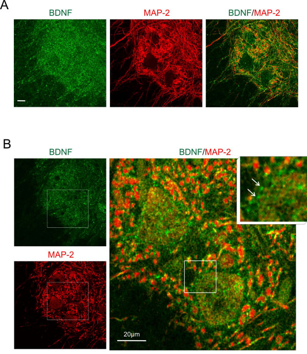
Figure 11.
Co-localization of BDNF (green) and dendritic MAP-2 (red) immunofluorescence in double stained cross-sections of the lumbar (L3/L4) segments in the intact spinal cord. A. Confocal images show the appearance of BDNF immunopositive and dendritic MAP-2 immunopositive structures in the ventral horn. Merged images (right) show regions of presumable co-localization of both proteins (yellow). The images are the stacks of twelve scans. Scale bar, 20 μm. B. The same area shown on the image of 1-μm-thick scan. The framed areas were enlarged and overlaid to reveal co-localization (right). Numerous MAP-2 positive dendritic profiles contain small, mostly perimembranous, BDNF accumulations. Scale bar, 20 μm. Inset: the arrows point to very small perisomatic BDNF immunofluorescent deposits which do not label for MAP-2.

