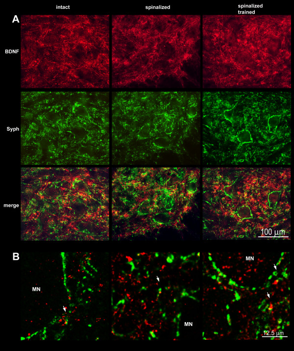
Figure 12.
Co-localization of BDNF (red) and synaptophysin (green) immunofluorescence in double stained cross-sections of lumbar (L3/L4) segments of the spinal cord in the intact (left column), spinal (middle column) and spinal trained (right column) rats. A. Two-color immunofluorescence photomicrographs demonstrate virtual lack of co-localization of BDNF (red) and synaptophysin (green) in the ventral quadrant of L3/L4 segment of the spinal cord in all experimental groups. Scale bar, 100 μm. B. Representative single confocal scan (1 μm thick), confirming rare co-localization of BDNF and synaptophysin (arrows) in neuropil of motor nuclei. MN- large neurons of lamina IX. Scale bar, 12.5 μm.

