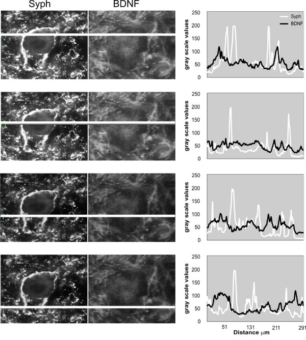
Figure 13.
Distribution of BDNF and synaptophysin immunofluorescence signal across a large neuron of lamina IX. Left panel: immunofluorescence signal, expressed as gray scale level, was measured in the narrow windows (white horizontal lines). Right panel: peaks of BDNF and synaptophysin signals overlay rarely indicating that these proteins often occupy different cellular compartments.

