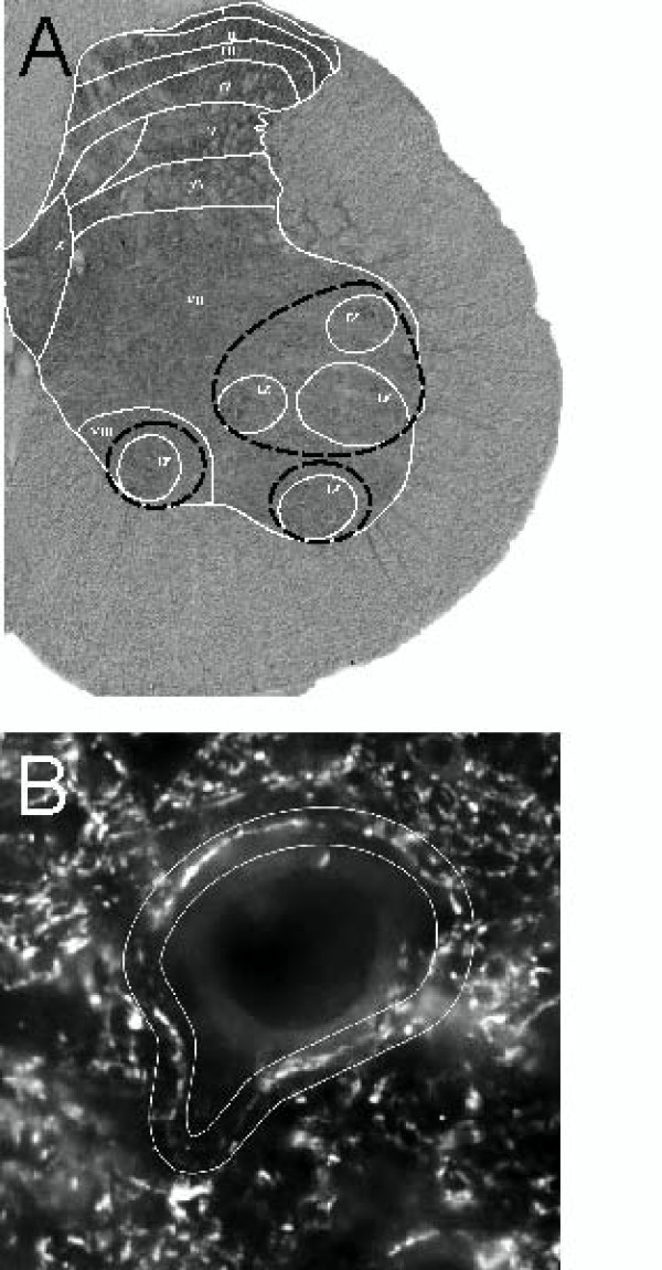
Figure 2.
Images of the spinal cord tissue sections with delineated areas which were subject to quantitative analysis. A. Brightfield photomicrograph showing immunocytochemical labeling of BDNF on a cross section of the lumbar (L3/L4) segment of the spinal cord in the intact rat. Spinal cord laminae distinguished in L4 segment [27] are overlaid. Areas covering motor nuclei (lamina IX) delineated with dashed black lines were taken for quantitative evaluation of BDNF immunoreactivity. B. Photomicrograph showing immunofluorescence of synaptophysin around a large neuron in the ventral horn of the lumbar (L3/L4) segment of the spinal cord. Delineated areas (white lines) surrounding large neurons of lamina IX were taken for quantitative estimation of the synaptophysin immunofluorescence intensity.

