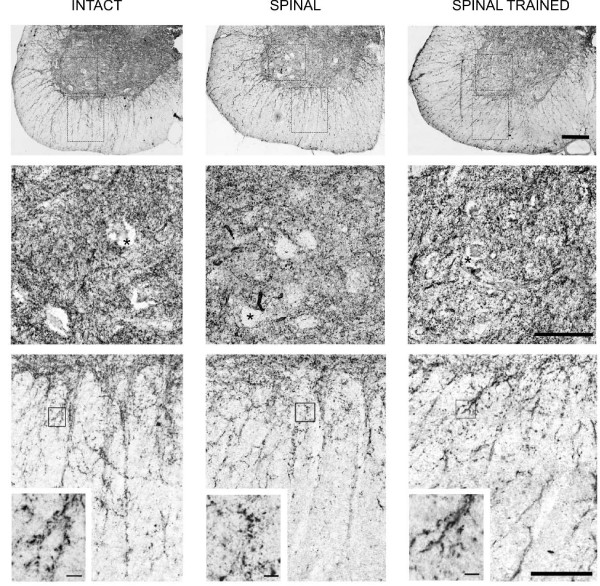
Figure 6.
Synaptic zinc staining pattern in the lumbar spinal cord of intact (left), spinal (middle), and spinal trained (right) rats. Upper panel: representative photomicrographs of 20-μm-thick cross-sections of ventral quadrant of the lumbar segments of the spinal cord. The framed areas of lamina IX are shown enlarged in the middle row and those sampling ventral funiculus are shown in the bottom row. Asterisks (middle row) mark large cell bodies which are devoid of the synaptic zinc-positive grains. Note that in the gray matter there are no visible differences in the synaptic zinc distribution between intact, spinal and spinal trained rats. Insets in the bottom row show, at high magnification, the bundles of processes radiating from the gray into the white matter demarcated by zinc-positive grains. This grainy encrustation seemed to be more prominent on fibers in the intact than in two other groups and was assessed quantitatively (see Figure 7). Scale bars: upper row, 250 μm; middle and bottom rows, 100 μm, insets, 10 μm.

