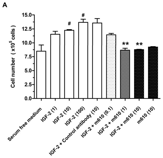
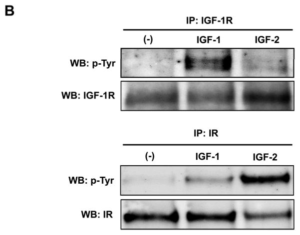
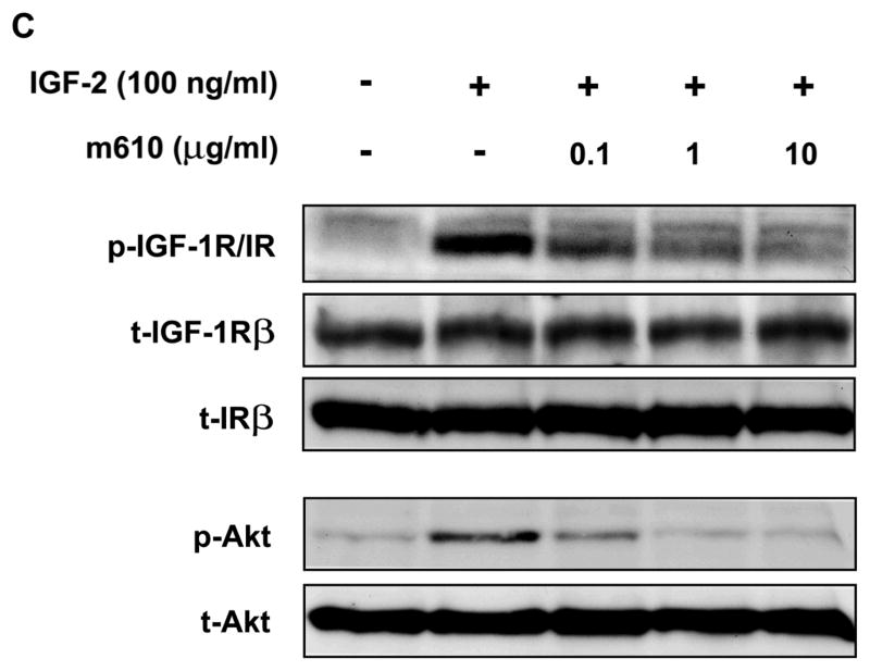
Fig. 6. M610 inhibits IGF-2-induced cell proliferation and phosphorylations of IGF-1R/IR and Akt in MDA PCa 2b cells.
A, MDA PCa 2b cells were treated for 48 hours with serum-free medium alone, with various concentrations of IGF-2, with the control antibody plus 100 ng/mL of IGF-2, or with various concentrations of m610 in the presence or absence of 100 ng/mL of IGF-2. The cells were counted using trypan blue dye exclusion. Data are the means of triplicate determinations and are representative of three independent experiments; bars, ± SE. #P < 0.05, compared with serum-free medium. **P < 0.01, compared with control antibody group. B, The cells were treated with 100 ng/ml of IGF-1 or IGF-2. Tyrosine phosphorylation of immunoprecipitated IGF-1R or IR were examined by a western blot analysis. WB, western blot; IP, immunoprecipitation; p-Tyr, phosphorylated tyrosine. C, The cells were pre-incubated with the indicated concentrations of m610, and 100 ng/mL of IGF-2 was added. IGF-1R/IR and Akt phosphorylation levels and the total protein levels in the lysates were examined by western blot analysis. p-, phosphorylated; t-, total.


